Nasal Valve Collapse Surgery - Before and After
- Dr. Waterman

- Oct 15, 2019
- 2 min read
The nasal valve helps keep the nostrils open especially when inhaling, and---as its name would suggest---nasal valve collapse occurs when one or both nostrils doesn't stay open. This can escalate and contribute to other sinus and nasal conditions, particularly regarding sinus drainage and infections of the nose and sinus.
Nasal valve collapse can occur internally or externally. Internal nasal valve collapse is recognizable when a person quickly sniffs through their nose and the nostril pulls inward near the alar crease, the fold between the nostril and the cheek. Internal nasal valve collapse can be treated with implants and other structural supports that help to keep the airway open.
An external nasal valve collapse is more apparent than an internal nasal valve collapse, and can be recognized by the rim of the nostril itself collapsing into the airway upon inhale. External nasal valve repairs are inherently more challenging to surgically correct because the structure of the nostril along it's rim is more delicate, more mobile, and of course exposed. Grafts are commonly required, along with precise reconstructive assembly of native cartilage, to provide a proper and more functional nostril.
As with surgery and treatments for other functional problems, realistic goals and expectations should be understood in terms of improvement by both the surgeon and the patient. While surgery can greatly reduce the severity of symptoms and improve aesthetics, conditions like nasal valve collapse can be persistent, and difficult to entirely eliminate.
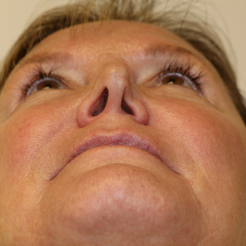
This patient was experiencing breathing issues and other functional problems due to an external nasal valve collapse. These photos are from before and 2-weeks after surgery. You will notice that one nostril is collapsed. This is an external nasal valve collapse because the compression is occurring at the alar rim, not above at the alar crease. In this case, a cartilage graft harvested from the patient's ear was used to provide the structural support necessary for proper function.
Just 2-weeks after surgery, this patient is already experiencing functional improvement, despite residual swelling. You can see that asymmetry has been greatly improved compared with the before- photos, but also how the nostrils are not perfectly identical mirror images. The procedure was a success, the patient was pleased with the improvement of both function and form.
Learn more about Ear Cartilage Grafting by reading our next blog post (coming soon!). Check us out on Google Maps!


